The clinical validation of a radiation-free technique, based on 3D tomographic ultrasound and supercomputing, has begun, which could revolutionize the diagnosis of breast cancer.
The European project QUSTom, which under the coordination of the Barcelona Supercomputing Center – Centro Nacional de Supercomputación (BSC-CNS) aims to introduce a new modality of medical imaging based on 3D tomographic ultrasounds and supercomputing, has started at the Vall d’Universidad Hospital. ‘Hebron of Barcelona clinical validation with patients for the early detection of breast cancer. This new technique could revolutionize the diagnosis of this type of tumors, since it is completely harmless for women and offers a much more complete image from a functional and multiparametric point of view.
In the coming weeks, volunteers will be recruited to participate in this initiative from among the women who participate in the Vall d’Hebron Hospital’s breast cancer early detection screening program. Unlike traditional ultrasound devices used in gynecology, which provide real-time images, this innovative technology prioritizes maximum image quality to improve diagnostic accuracy. It not only aims to complement and improve the diagnosis of breast cancer, but also potentially replace current diagnostic methods, such as mammograms, which use x-rays.
A computerized tomograph unique in the world
To take images, a 3D Ultrasound Computerized Tomography (3D USCT III) will be used, designed and built by the
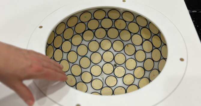
Karlsruhe Institute of Technology (KIT) in Germany, one of QUSTom’s partners. It is the only complete device of these characteristics in the world. With a 3D hemispherical aperture consisting of 2,304 individual transducers, acting as transmitters and receivers, it is used to examine breast tissue for pathological changes. The KIT has been working on the development of additional prototypes, but the first to undergo validation with patients is the one currently in Barcelona.
Before the device reached its current stage, it passed a series of electrical and ultrasound safety tests supervised by a certified medical device testing laboratory in Germany. Once all the data has been collected, it will be reconstructed using the 3D full-wave inversion algorithm and transformed into high-resolution medical images using the power of the MareNostrum5 supercomputer, at the BSC, with UBIware software developed by FrontWave Imaging, a company whose creation has been promoted by the BSC, and Imperial College London in the United Kingdom, which is also the sponsor of the clinical validation. The project also incorporates concepts such as multimodal imaging and true 3D imaging, representing an unprecedented combination in breast imaging using ultrasound.
Digital twin of simulated breast tissue in MareNostrum 5
Using the MareNostrum 5 supercomputer, around 50,000 simulations of ultrasound waves will be performed for each reconstructed image. In 2D this problem is not very challenging and can be computed in a few Graphics Processing Units (GPUs), in a conventional cloud. In 3D, however, the problem becomes gigantic, so much so that until today no one has applied the best image reconstruction techniques through simulation to 3D data such as those that will be used in this clinical validation. “We will be pioneers in this thanks to the use of MareNostrum 5. What we can achieve in a few days at the BSC would take years on a normal computer,” said Josep de la Puente, BSC researcher and coordinator of QUSTom.
The BSC researcher explains that, at its core, the project builds a digital twin of the breast tissue and the ultrasound measurement device. This digital twin replicates any ultrasound emission emitted by the physical device used by the radiologist. “Consequently, we can acquire not only a post-processing image, but a complete three-dimensional map that details the properties of the tissue in each pixel,” said De la Puente.
“This new diagnostic tool will allow us to offer a more complete image from a functional and multiparametric point of view, avoiding the use of ionizing radiation and improving the comfort of women during their annual radiological examination, in order to detect breast cancer early,” highlighted Ana María Rodríguez Arana, head of the Women’s Radiology Service at the Vall d’Hebron Hospital and principal investigator of the Molecular Medical Imaging group at the Vall d’Hebron Research Institute (VHIR).
Harmless to women
Unlike other tests such as mammograms, QUSTom’s technology does not use radiation. The new device offers potentially higher image quality and better tumor tracking through the use of ultrasound and supercomputing. The exam is painless and more comfortable for the patient. The technology has wide application, but may be particularly beneficial for people with dense breast tissue, which represents 40% of women worldwide, according to the Spanish Society of Senology and Breast Pathology (SESPM).
For the development of the project, the BSC has used its experience in the detection and analysis of data obtained in mechanical wave problems, so that the algorithms used to obtain medical images are inspired by other algorithms used in completely different research areas. , such as the analysis of the earth’s subsoil.
How does it work
The patient is placed face down on a bed, while her breast is immersed in a container filled with water at a temperature of 36.5 degrees Celsius.
Ultrasound is then used to take data from each breast separately.
The recorded data is transferred to a computer.
The procedure lasts approximately 3 minutes per breast.
In a matter of hours and after thousands of simulations, the software used in the supercomputer generates real, high-quality 3D images, capable of providing a more precise diagnosis. These images are ready to be analyzed by doctors.
QUSTom project technology. (Photo: Vall d’Hebron University Hospital)
Breast cancer: the most diagnosed type of tumor in the world
Statistics from the Spanish Society of Medical Oncology (SEOM) show that breast cancer is one of the most common tumors worldwide, with 2.3 million women diagnosed in 2020 and 700,000 deaths due to this disease during the same year. .
In Spain, approximately 36,395 new cases of breast cancer are expected to be registered in 2024, according to data from REDECAN, which represents a slight increase compared to the previous year.
Early detection of the disease plays a critical role as it can significantly increase survival rates. Although mammography is a widely used tool for the detection of breast cancer and has contributed to saving numerous lives, it is interesting to have a range of options for new non-irradiating technologies that can be used for diagnosis. (Source: Vall d’Hebron University Hospital)