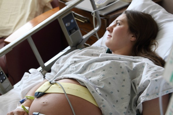
Women with poorly controlled diabetes multiply the risk of having a newborn with congenital malformations, making timely detection and treatment essential.
|
Summary:
Diabetes mellitus is increasing worldwide. Many people living with diabetes are unaware of the diagnosis, including women of childbearing age. Clinicians must manage current knowledge about congenital anomalies associated with maternal diabetes and be able to identify mothers at risk of having offspring with congenital disorders.
|
INTRODUCTION
Diabetes mellitus (DM) is one of the most common chronic diseases that affect humans.1 There are many different forms of diabetes, with different etiologies and clinical manifestations, the common feature among them being hyperglycemia.2 In 2019, approximately 15.8% of women experienced some degree of hyperglycemia during pregnancy; of them, 83.6% were due to gestational diabetes mellitus (GDM), 7.9% to pregestational diabetes and 8.5% to diabetes diagnosed for the first time during pregnancy.1
Women with poorly controlled diabetes in early pregnancy increase the risk of giving birth to a newborn with a congenital malformation by nine.3 The rate of congenital malformations in women affected by established type 1 DM is identical to that of those with established type 2 DM.4 While this is true, the association of congenital anomalies in the offspring of those with maternal GDM remains unclear. 4, 5, 6 Additionally, diabetes is more likely to cause miscarriage or stillbirth.7
MECHANISMS
Multiple mechanisms are involved in the pathogenesis of diabetes-induced birth defects.
Being the hyperglycemia The unifying characteristic of the different forms of DM, embryonic and fetal exposure to high concentrations of glucose is a major teratogenic factor.8 Hemoglobin A1c levels used as an indicator of glycemic control in diabetic women have been shown to correlate with rates of congenital anomalies.9 The crucial exposure time for the development of congenital anomalies in babies of diabetic mothers is during organogenesis, which occurs between weeks 2 and 8 of gestation.10
Even brief periconceptional exposure to hyperglycemia is sufficient to undermine embryonic development.eleven Baack et al., in an animal study, demonstrated that even in the absence of DM, hyperglycemia was capable of causing congenital defects in offspring.12 It has been shown that a high concentration of glucose is capable of altering the DNA methylation of crucial genes involved in embryonic development.13
Li and collaborators showed that advanced glycation end products (AGEs), whose production is accelerated under conditions of hyperglycemia, are capable of inducing congenital defects even in the absence of hyperglycemia itself.14 Experimental DM in animal models has been shown to be associated with overproduction of reactive oxygen species (ROS) and disruption of antioxidant defense.fifteen
Hyperglycemia promotes the entry of glucose into embryonic cells and, as a result, mitochondria receive a metabolic overload that results in increased ROS formation.16 Excessive oxidative stress then leads to DNA damage, microRNA disruption, and disruption of various signaling pathways, all of which result in aberrant organ development.17,18, 19, 20, 21
Laboratory experiments have shown that birth defects can be reversed by treatment with antioxidants such as N-acetylcysteine, resveratrol, and melatonin.22,23, 24 Furthermore, hyperglycemia has been shown to disrupt intracellular calcium homeostasis, leading to perturbation of organelle function and consequent increase in apoptosis. 25
CENTRAL NERVOUS SYSTEM
Maternal diabetes is known to disrupt the development of the central nervous system and cause birth defects.13, 26, 27, 28, 29
The central nervous system is among the systems commonly affected in a baby born to a diabetic mother. The most common birth defects are neural tube defects, holoprosencephaly and hydrocephalus.30, 31
Other central nervous system defects associated with maternal diabetes are schizencephaly, agenesis of the corpus callosum, and hamartomas.32, 33, 34, 35 Neural tube defects are a heterogeneous group of deformities characterized by an opening in the spine or skull, resulting from a failure in the normal closure of the neural tube during early embryonic development.36, 37
The anencephaly It is characterized by the absence of major portions of the skull and brain as a result of failure to close the rostral neuropore. 38 Encephalocele is a defect consisting of a sac-shaped herniation of neural tissue through an opening in the skull.39
The exencephaly It is characterized by the complete or partial absence of the bones of the roof of the skull with a properly formed brain.
The spina bifida It occurs when the vertebral column is divided (bifid) as a result of failure of the embryonic neural tube to close at the posterior end during the fourth week of embryonic development. It can manifest as meningocele, myelomeningocele or occult spina bifida.37
The holoprosencephaly It is an anomaly of brain formation in which a total or partial absence of division into hemispheres is observed. Symptoms of this condition include abnormal facial formation with the development of cyclopia, proboscis nose (or no nose), cleft lip and palate.40
The syntelencephaly It is a rare anomaly in which forebrain cleavage is absent in the region of the posterior parts of the frontal and parietal lobes, with normal interhemispheric cleavage observed anterior and posterior to the affected region.41
CARDIOVASCULAR SYSTEM
The most common congenital heart defects associated with maternal diabetes include patent truncus arteriosus, atrioventricular septal defect, heterotaxy, and single ventricle complex.42
Cardiogenesis is an elaborate process involving numerous types of tissues. Causal mechanisms of diabetes-induced heart defects include dysregulation of hypoxia-inducible factor-1, notch signaling, wingless-related integration signaling, and transforming growth factor-b signaling pathways.18, 43, 44, 45, 46
He persistent arterial trunk It is a defect that presents a ventricular septal defect (VSD), a single truncal valve and a single ventricular outflow tract. Pulmonary venous blood mixes with systemic venous blood at the level of the VSD and is then ejected into the single great artery.4, 48 The defect arises from incorrect creation of the conotruncal septal wall, and the common truncal root does not separate into aortic and pulmonary outflow tracts.49
He atrioventricular septal defectalso know as “atrioventricular canal defect”is a heart defect characterized by a varied level of atrial and ventricular septal defects along with a single or partially divided atrioventricular orifice.fifty This defect is the result of failure of fusion of the endocardial cushions. These cushions constitute paired bulbous mesenchymal structures (upper and lower) that develop early in embryogenesis at the atrioventricular junction. They develop towards each other and finally fuse after 4 weeks of development to form a continuous septum and subsequently form the valves. Lack of fusion results in different degrees of atrioventricular septal defects.51, 52 It has been shown that embryos from diabetic pregnancies have poor development of the endocardial cushions.53, 54
Heterotaxy is a broad term covering various conditions characterized by an abnormal arrangement of the thoracic and abdominal organs along the left-right axis of the heart that cannot be described as situs inversus.55, 56 There is a wide variety of lesions of the cardiac structure that can involve atrial anomalies, conotruncal anomalies, atrioventricular canal defects, total and partial anomalous venous return, and ventricular outflow abnormalities.57
Systemic venous abnormalities and various arrhythmias may also be present.57, 58
He single ventricular complex It is a combination of congenital heart defects in which 1 of the ventricles is substantially underdeveloped or the interventricular septum has not formed.59 This defect originates during the first 8 weeks of development.60
Other congenital heart defects less frequently associated with diabetes include tetralogy of Fallot, D-transposition of the great arteries, double outlet of the right ventricle, anomalous pulmonary venous return, coarctation of the aorta, and aortopulmonary window.: 42, 61 Animal data have also shown that pregestational diabetes can impair the development of the coronary vasculature and cause hypoplasia of the coronary arteries.twenty
CRANIOFACIAL AREA
Craniofacial defects found in children of diabetic mothers include orofacial clefts, oculo-auriculo-vertebral disorder (OAVD) spectrum, anophthalmia and microphthalmia, cataracts, coloboma, choanal atresia, and others. 5, 62
The orofacial clefts They develop as a consequence of aberrant embryonic development of the nasomaxillary complex. They can be defined by the absence of continuity of the palate, the upper region, alveolar margins and upper lip. The defects can be of various degrees and lateralities.63, 64 Occasionally, cleft defects can involve various other bony and soft tissue structures of the face.65 Such defects have also been associated with maternal DM.66
The spectrum of oculo-auriculo-vertebral disorder (OAVD) includes a heterogeneous group of disorders characterized by abnormalities of the structures derived from the first and second pharyngeal arches during embryogenesis.67 These disorders, as their name suggests, are characterized by abnormalities of the ear, eyes, and spine.
The spectrum is subdivided into 3 disorders: OAVD, hemifacial macrosomia, and Goldenhar syndrome.
Associated clinical features also include brain abnormalities and developmental delay.67 Considering that most abnormalities are found in structures derived from neural crest cells, it has been hypothesized that poorly controlled maternal diabetes disturbs the cephalad migration of neural crest cells.69 Furthermore, it has been suggested that the cause could be the alteration of the Pax3 gene pathway due to hyperglycemia and the subsequent oxidative stress.70