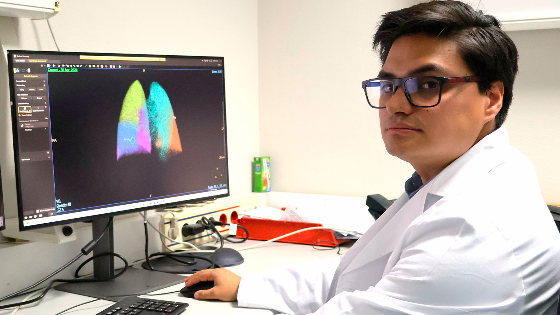

radiologist at the Nuestra Señora del Rosario University Hospital
Artificial intelligence is already decisive in the field of radiodiagnosis by simplifying processes, increasing efficiency in reading studies or increasing diagnostic precision.

Artificial Intelligence (AI) has become, in recent times, an essential ally of medical specialists. In fact, in the field of radiodiagnosis has been part of it for a long time, although It is now when it is most decisiveby simplifying processes, increasing efficiency in reading studies or increase diagnostic accuracy.
At the Nuestra Señora del Rosario University Hospital in Madrid, it is used for early diagnosis of lung and prostate cancers. Dr. Nicolás Almeida, one of the specialists in the service directed by Dr. Eliseo Vañó, explains to OKSALUD the important contributions of AI in the daily work of radiologists: “AI is destined to stay to improve our work and, above all, benefit the health of our patients.”
QUESTION.- When does AI break into the field of radiodiagnosis?
ANSWER.- AI has been part of radiology for quite some time, but it is in recent years that very rapid progress has occurred and it has definitively emerged as a common element in our field. In the beginning, the first AI-based diagnostic methods had lower performance. However, several recent AI models are capable of equaling and even surpassing humans in certain diagnostic processes. A summarized way of the diagnostic process in radiology would be: detect a finding, describe it, characterize it, reach a diagnosis and, subsequently, monitor the disease. The effectiveness of this process will fundamentally depend on the experience and knowledge of the radiologist and will sometimes be subjective. AI excels at recognizing complex patterns in images, data and can provide quantitative evaluation in an automated manner. This increases the precision and reproducibility of the scans.
Q.- Ally or enemy of the medical professional?
A.- AI is not an enemy of the radiologist, rather it is his main ally, since it simplifies various processes, increases efficiency in reading studies and increases reproducibility and diagnostic precision. AI helps the radiologist and the patient, as it facilitates the detection and adequate characterization of the findings, offering a clear health benefit. All this without removing the human side of clinical practice.
Q.- When did you join the Nuestra Señora del Rosario University Hospital?
A.- AI has been part of our service almost from the beginning and continues to do so in our usual practice. For example, in the quantification of coronary calcium load, segmentation of arteries, quantification of vascular stenosis, characterization of atheromatous plaques, among others. However, we must highlight that since the service acquired the latest generation CT 7500 spectral CT equipment, the application of AI has been enhanced and new opportunities for its application have emerged.
Q.- What applications does AI have in CT or MRI scans? What are your main contributions?
A.- Countless. In the case of CT, they can be specified in detection of pulmonary nodules, automatic segmentation of organs, quantification of vascular stenosis, characterization of atheromatous plaque, estimation of the probability of myocardial ischemia, tissue characterization, quantification of iodine concentration , decrease in contrast dose among others. If we talk about magnetic resonance: reduction in image acquisition times, tissue characterization, cardiac segmentation, cerebral perfusion, assessment of myocardial deformation, among other benefits.
Q.- What advantages does it generate for the radiologist? And to the patient?
A.- For the radiologist, it reduces the time spent interpreting the studies, facilitates interpretation, increases reproducibility, favors the monitoring of pathologies, allows findings to be quantified and increases diagnostic precision. For the patient, this means less waiting time for the results of their tests and reduced diagnostic uncertainty, among other advantages.
Q.- Is a new path full of opportunities opening up from now on?
A.- Definitely yes. In our service we are regularly using AI for the early detection of prostate cancer and lung cancer. Regarding prostate cancer, it is the second most common in men, and it is estimated that one in eight will be diagnosed with this disease. MRIs are now the standard of care for these patients. In fact, at the Hospital we do about 600 a year. Now, the recent acquisition of the QP-Prostate software, developed by the Quibim company, makes it possible to detect prostate cancer in its initial stage, significantly improving the prognosis and subsequent treatment of the disease. This technology enables advanced analysis of MR images, highlighting its ability to identify early signs of cancer with unprecedented precision, facilitating faster evaluation that contributes to the personalization of treatment for each patient.
Q.- And what about lung cancer?
A.- Regarding lung cancer, we must not lose sight of the fact that it is responsible for 21% of all cancer-related deaths. Of all diagnosed cases, only 20-30% are diagnosed in early stages. This is a fact that influences the prognosis of the disease, because the 5-year survival rate in early stages is 85%, while for advanced stages it is only 10%. Most cases are asymptomatic and when symptoms appear, the disease is usually very advanced.
The early detection program for lung cancer is based on performing a low-dose radiation chest computed tomography, which allows the assessment of the lung parenchyma, its fundamental objective being to detect suspicious lung nodules in order to diagnose lung cancer early. Screening has shown in some studies a decrease in specific mortality for lung cancer of up to 25%.
The scans are performed every 12 months and different monitoring or therapeutic strategies are adopted depending on the findings.
In our service we have recently started the lung cancer early detection program and for this we use artificial intelligence software for automated nodule detection as a second reading (Philips Lung Nodule Assessment based on ClearRead CT). In this way, we carry out a first interpretation of the study in the usual way and with the AI software we proceed to a second reading to detect nodules that may have been omitted. In addition, this program allows you to perform measurements quickly, obtaining axial diameters and volumes accurately.
The software takes between one and two minutes to read the study, segment the lungs and detect nodules automatically, providing all its measurements. In addition, it offers the opportunity to generate an automatic structured report. All of these factors help the radiologist in the interpretation of these studies, increasing diagnostic precision and reducing reading time.
But this is only the first step in this field. AI in lung cancer in the future will allow, based on its radiological characteristics, to estimate the histological type, the response to specific types of treatment and, probably, the prognosis.